
| URSA 200 is the first
microanalyser to completely integrate digital electron and spectral imaging with
the x-ray analyser. Not only image acquisition software and spectrum acquisition
software are parts of a single microanalysis application, but all the microscope
beam control and spectrum acquisition circuits reside on the same board . This
ensures there are no delays in switching from one mode of operation to another,
no extra PC slots required and no cabling for often troublesome connections
between separate modules.

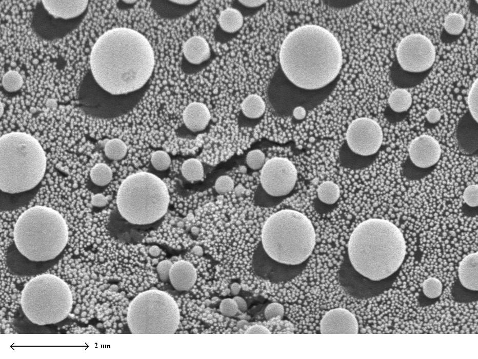
Despite the very straightforward user interface, URSA 200 packs
some very powerful features. High definition electron images from two
simultaneous sources (typically secondary and backscattered electron detectors)
can be acquired in seconds at resolutions of up to 2048 and with 4096 grey
levels. Thanks to the unique pixel integration technique used by URSA 200,
the quality of the image rivals that of the microscope. Even mains
synchronization can be employed for those low kV, high magnification images.
|
(click on the image to enlarge) |

|
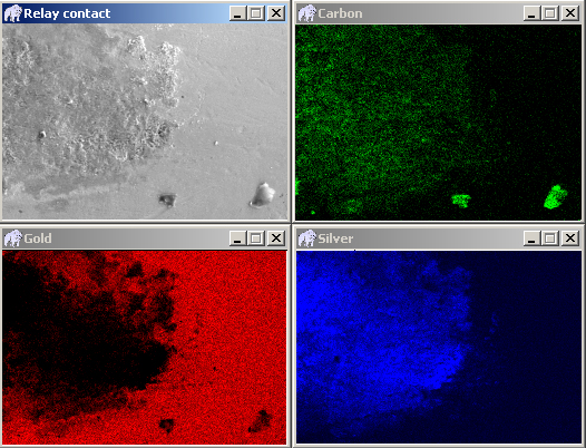
URSA 200 takes full advantage
of its high throughput capability by acquiring full spectrum information from
each pixel as it scans the sample digitally with dead time corrected dwell time.
Any number of element maps (together with two electron images) can be viewed
during acquisition. More importantly, maps of additional elements can be
examined after the spectral image file has been acquired. Special software tools
can automatically reveal all the elements contained in the spectral image file
and even produce maps based on chemical composition. URSA 200 also
allows you to extract aggregate spectra from user defined areas on the image.
Any areas can be defined, whether by marking a rectangle, a polygon or using the
free hand drawing tool. Naturally you can also extract elemental line profiles
along lines drawn in any direction.
|
(click on the image to enlarge) |
URSA 200 beam control also allows you to position the beam in spot
mode for spot analysis and can be programmed to sequentially acquire spectra
from a series of pre-selected spots for unattended operation. It can also drive
microscope beam along a user defined line at any orientation to provide live
line profiles.

All these modes of operation share a unified user interface that supports many
powerful features. All images are acompanied by a calibrated micron bar and can
be zoomed or panned to any size. A linear measurement tool is always available.
Many image processing functions such as pseudo coloring, histogram equalization,
contrast and brightness control, smoothing and image annotation can be performed
with a click of a button. Intensity histograms as well as image statistics
display (minimum, maximum, average, S.D.) are also just a mouse click away.
For users doing high resolution microanalysis over extended periods of time, URSA
200 offers a stage drift correction option. With this option, image
drift is automatically detected by the software, and corrective signals are sent
to the microscope scanning coils to return the image to its original position.
To handle the vast amount of information acquired in the spectral image, URSA
200 employs a unique on-the-fly compression technique. This keeps URSA's
run-time memory consumption to a minimum, and ensures saved files are compact.
Images can also be exported to the popular TIF format, or pasted into other
applications. URSA reports allows the creation of report templates,
where images (as well as spectra, quant results, and any other available
information) are automatically placed into custom URSA reports.